


Optical Microscopy Application: Differential Interference Contrast
저자: Stephan Briggs
Differential Interference Contrast (DIC) is a polarization technique in optical microscopy that uses a polarizer, analyzer, and additional polarization optics such as a Nomarski or Wollaston prism. In simple DIC setups, the only required components are a polarizer and an analyzer. The polarizer is typically positioned below the specimen, and the analyzer above the objective lens. The polarization axis of each can be rotated to adjust contrast and throughput, but the two components typically act with respect to one another.
Introducing a Nomarski or Wollaston prism into a DIC optical microscopy setup separates polarized light into two rays that are polarized at 90° to one another. The focal point of the prisms rests outside the glass element, and allows active focusing in the upright DIC microscope system. A second prism is required to join the two light rays back together at a polarization of 135°.
Image Appearance
Figure 1 is a darkfield illumination image with specialized polarized techniques applied to it. The tissue fibers exhibit added complexity, detail, and texture as the light scattered by it is controlled and captured differently by its state of polarization. The image shows details about absorption color, optical path boundaries, and refractive indices, along with whether or not a sample is isotropic and anisotropic. Polarization techniques in optical microscopy such as DIC are invaluable in the identification of unknown samples that exhibit birefringence.
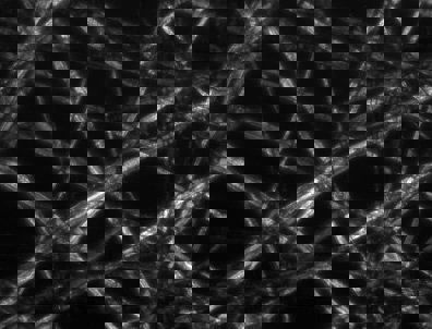
Figure 1: Polarization-Contrast Image of Tissue Paper
Technical Details
Figure 1 was captured using a simple DIC microscope. From bottom to top, there are five key assemblies and components.
- Light source emits unpolarized light into microscope and is immediately oriented to 45° by the first polarizing filter.
- Polarized light enters Wollaston prism and is separated into two rays at 90° to one another.
- Both rays are focused by condenser lens to sample at specific points.
- Rays propagate through sample at a separation point which mimics the resolution of the microscope (0.5μm resolution, rays focused at 0.5μm separation). Light is collected by objective lens and focused to second Wollaston prism.
- Second Wollaston prism combines two rays into one polarized ray at 135°, which results in improved contrast in the sample image: improvements in bright spots, dark spots, and overall focus.

Figure 2: Optical Path of Differential Interference Contrast Microscope
Additional optical microscopy applications include brightfield illumination, darkfield illumination, phase contrast, and fluorescence.


본사 및 지사별 연락처 확인하기
견적 요청 도구
재고 번호 입력 필요
Copyright 2023, 에드몬드옵틱스코리아 사업자 등록번호: 110-81-74657 | 대표이사: 이준호 | 통신판매업 신고번호: 제 2022-서울마포-0965호, 서울특별시 마포구 월드컵북로 21, 7층 (서교동, 풍성빌딩)
The FUTURE Depends On Optics®